
Mandible and Temporomandibular Disorder (TMD)

Did you know that 18 hours after a tooth extraction, your mandible (specifically the condyle of the mandible) shifts its shape to accommodate the change?
And did you know that because the joint linking the mandible with your skull — the temporomandibular joint (TMJ) — is located so close to the ear canal, the tension in the jaw might affect your hearing?
Our mandible, or lower jaw if you like, is the largest and strongest bone in the human skull, built with strong diploic bone. It’s also the bone that enjoys the most movement within the skull. It is attached to the skull by very strong muscles and ligaments, allowing us to open our mouth wide to bite an apple, chew it, swallow it, and also speak about it later. Once we finished with the apple, we can use the mandible for kissing and singing too :-)
The mandible holds our lower teeth in place and it is so strong that you would need a force of at least five hundred pounds to break it. With such impressive tensile strength, it weighs only slightly more than an ounce.
Mandibular development

The Mandible starts developing when we are breastfed. Alternating between the right and left breast allows for equal development of the TMJ and the atlanto-occipital joint, i.e., the joint between your skull and the top vertebra of your neck. When babies are bottle-fed, it’s really important to switch sides from left to right to assist the equal development of the TMJ and the baby’s neck.
Scar tissue in the mandible
Think of how many visits to the dentist you’ve had. Some of them might have left scars and marks on your mandible that have never been treated. Just like with any other surgery, the scars in the mandible shouldn’t be left unattended. In the ideal word that I keep dreaming of, each visit to the dentist would be followed by an appointment with a craniosacral therapist, so that there are no scars and tensions that are left untreated.
The mandible has enough to deal with without dental work. It is often linked with emotional stress and trauma. Think of the last time you found yourself feeling a real rage, totally gripped by it. Your mouth was likely to be so tight that you could hardly open it. Or perhaps, bring your mind to the last time you were nervous before a public speaking event. Do you remember how you tried to overcome the tightness in your mouth and clenching of your teeth? A lot of us never get relief from stress like this — we just get used to it. And it isn’t a trivial amount of tension, either. The jaw can bench-press 180 pounds per square inch! Ongoing tension in the mandible can lead to grinding the teeth, headaches, gradual degeneration of the TMJ joint, cervical spine arthritis, visual problems and hearing loss.
Mandibular fascial connections
In the Anatomy Trains model of understanding the human body, the mandibular fascia belongs to the Deep Front Line - that’s the myofascial core of our being. The deepest layer of ‘us’ that extends from the underside of the feet, travelling up via the deep compartment of the lower legs, behind the knee and towards the inner thigh, continuing via the lumbar spine, hip flexors, abdominal muscles, diaphragm, ribcage, thoracic viscera, and finally completing the journey in the underside of both the neuro- and viscero-cranium, which is what we are taking about here.
It’s fascinating to me that the mandible is closely connected to the movement of the feet. Whilst stepping forward with our foot, our TMJ responds by rotating open on the side of the foot that is making the movement. Pretty cool, no?
Mandibular Movement
I mentioned earlier that the mandible enjoys the most movement out of all of the bones of the human skull. So what are these movements exactly, and what would we evaluate when testing the mandible? There are eight possible movements to be aware of:

Protrusion - anterior glide
Left rotation
Right rotation
Left lateral shear
Right lateral shear
Retrusion (retraction) - posterior glide
Decompression
Compression (this last movement is the only one that we wouldn’t use for clients with dysfunction of the mandible)
The primary axis of movement of the mandible is located at the base of dens — that’s the structure at the top of the second vertebra of the neck, where the first vertebra and your skull turn — a very tricky place to access! I should know: I have tried it to access it on my poor colleagues during training (kids, don’t try this at home: we were supervised and guided by a very experienced teacher at the time). The secondary axis of motion is around the TMJ.
Temporomandibular Joint (TMJ)
The TMJ — the joint that connects our mandible with the temporal bone of the skull — is the most busy and frequently used joint in the body, moving on average 2,000-3,000 times per day. The joint is equipped with an articular disc, an S-shaped structure also known as a saddle joint, which cushions the space between the two bony surfaces and prevents the deterioration of the bones. Long-term tension of the surrounding muscles can actually lead to gradual dislocation within the joint space whereby the cushioning of the disk is no longer able to prevent the damage.
Temporomandibular Disorder (TMD)
We don’t usually learn about the TMJ unless we are diagnosed with temporomandibular disorder (TMD). The first sign of a problem might be clicking of the jaw. If the tension in the jaw is not released and persists for a long time, it might develop into chronic pain. It feels like the worst toothache that you can imagine, and it just doesn’t go away.

MUSCLES THAT MIGHT BE INVOLVED IN TMD
There are 16 muscles attaching directly to our mandible (only the shoulder joint has more - 17) and … wait for it…136 muscles connecting to it. Many of them might need treatment to relieve pain in the TMJ region. I will only describe the main muscles here (I can almost hear you sighing with relief right now :-)
Masseter

Used primarily for chewing, the masseter is the strongest muscle in the body in relation to its size. If all the muscles of the jaw work together, the masseter can close the jaw with a force of 200 pounds at the molars. It’s enough to bite off a finger! This muscle elevates our lower jaw and is also used for speaking, swallowing, and clenching the jaw, whether due to acute or long-term stress. It is square shaped and composed of two bellies - the superficial belly can be accessed and treated by touching the face, and the deep belly can be accessed and treated with intra-oral work. It’s a major pattern-setter for mandibular movement, and hence very important part of assessment and treatment of any jaw dysfunction or pain. The masseter originates in the maxilla (upper jaw bone), zygomatic arch and temporal bone. It inserts into the angle and ramus of the mandible. It is innervated by the mandibular division of the trigeminal nerve.
Temporalis

The temporalis muscle can be seen here as a fan shaped structure above the ear.
The temporalis muscle is what we usually find ourselves massaging on the side of our heads when we have a headache. In fact, tension in the posterior part of the muscle may cause TMJ pain and headaches. It’s located on the side of the skull, originating from the temporal fossa and fascia (spanning across frontal, temporal, parietal and sphenoid bones). This big muscle travels down and dives under the zygomatic arch to insert into the hook-like structure of the coronoid process of the mandible. Similarly to the masseter muscle, it elevates our jaw, and is innervated by the mandibular division of the trigeminal nerve.
Medial and Lateral Pterygoids
These two muscles act as assistants to the functions of the masseter and temporalis. The medial pterygoid helps to elevate the mandible and the lateral one helps with protraction, i.e., moving the jaw forward.
The medial pterygoid is the identical twin of the masseter muscle. The masseter is located on the outer side of the mandible and the medial pterygoid is situated on the inner side. Together they create a V-shape structure. The medial pterygoid has two origin points. The first one is at the medial surface of the lateral pterygoid plate of the sphenoid bone, and the second one is at the tuberosity of the maxilla (the upper jaw). The muscle inserts into the medial surface of the ramus of the mandible and is innervated by the trigeminal nerve.
The lateral pterygoid also has two bellies, therefore two origin points, both located on the sphenoid bone. The first origin point is the infratemporal surface and crest of the greater wing of the sphenoid. The second one is the lateral pterygoid plate of the sphenoid bone. They both insert into the articular disk and capsule of the TMJ and the neck of the mandible. Like all the muscles mentioned above, this one is also innervated by the trigeminal nerve.
When the muscles of the jaw are tight and compressed, they can exert pressure of around 180 pounds on our teeth. When healthy, however, the muscles will exert no pressure at all and keep the articular disc of the TMJ protected and working perfectly. Due to the anatomical differences between female and male bodies (more negative pressure when opening the jaw in women) 70% of people who suffer from TMD are women.
If the jaw seems to be retracted - (pushed backwards) we would look at the muscles forming the ‘floor of the mouth’:
1. Mylohyoid
2. Digastric
3. Stylohyoid
4. Geniohyoid
Mandibular ligaments
Whilst determining the course of treatment, we might also evaluate the four ligaments of the mandible:
Stylomandibular ligament - connects the styloid process of the temporal bone with the mandible
Temporomandibular ligament - links the temporal bone with the mandible
Sphenomandibular ligament - shaped like an Eiffel tower, this ligamentous structure links the sphenoid bone with the mandible
Retrodiscal ligament - a thickening of the TMJ capsule
TREATMENT CONSIDERATIONS
We would check for mandibular tensions when treating:
TMD
Any type of headache (because of the mandible’s connection with the temporal and sphenoid bones), i.e., cluster headache, tension headache, menstrual headache, sinus and eye pain, migraine
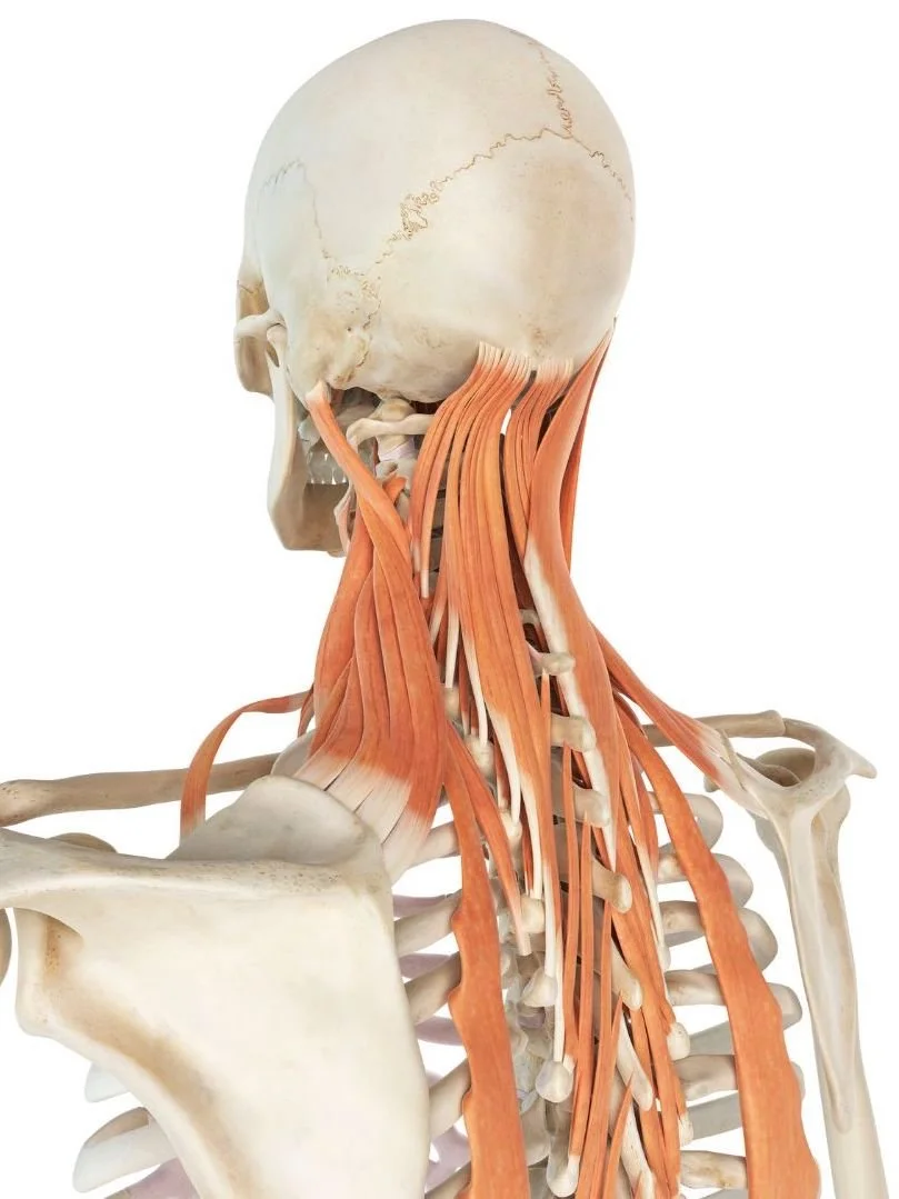
Neck pain (especially the back of the neck and discomfort coming from tension around the first three cervical vertebrae). It is impossible to evaluate the mandible without looking at and assessing the structures and possible tensions in the neck. The neck muscles, especially the posterior group, will likely be involved in the pain pattern associated with TMD. It works the other way around, too: it is impossible to release the neck fully without checking for possible tensions in the mandible.
Breathing difficulties - we would also consider releasing the mandible to improve breathing in asthmatic patients and other people presenting with breathing problems
Osteoarthritis
Lower back pain
Sacral and pelvic pain or blockages - because of the neurological and energetic connection between the mouth and the perineum, and the symphysis mantis and symphysis pubis
work with trauma, aggression, suppressed emotions, fear of speaking out
Resources:
Andrew Biel, Trail Guide to the Body, 2013
Frank Netter, Atlas of Human Anatomy, 2015
Hugh Milne, The Heart of Listening. The Visionary Approach to Craniosacral Work, 1995
Tom W. Myers, Anatomy Trains. Myofascial Meridians for Manual & Movement Therapists, 2014